Detailed Eye Imaging
Detailed Eye Imaging namely Optical CT is a powerful tool used in establishing precise diagnosis.
Below we list few features of the equipment and it’s diagnostic capabilities. Back of the Eyes.
-
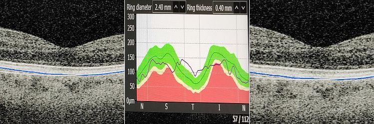
To appreciate the imaging capability of the scanner please view the supplied images. The capability helps our Optometry Doctors differentiate between various pathologies and manage the patients accordingly.
With respect to imaging the back of the eyes , we recommend 4 phase differentiation management algorithm protocols for the practice. These breakdown into 4 categories;
*pathology which can be managed pharmacologically with NSAIDS (inhouse)
-
- pathology which requires intervention with specific agents as; Anti Vaso-Endothelial Growth factor inhibitors (anti-veg F ) or Ocriplasmin, or other more specialized pharmacological agents which is done via eye surgeons.
-
- vitero-retinal surgical intervention, or medical laser (specialized surgery )
-
- pathology requiring general systemic approach ie autoimmune conditions, metabolic disorders, general health management (General Medical Practitioners)
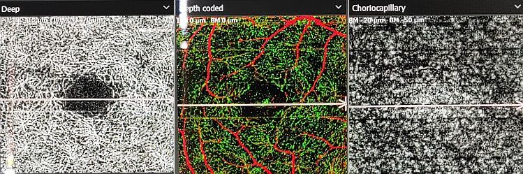
Angiographic Images.
Posterior imaging of vessels. Images show anterior retinal vascular plexus ,deeper retinal ,and features of choriocapillary network.
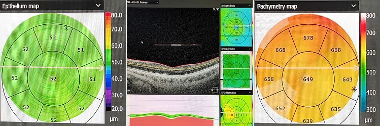
Front of the Eyes.
With respect to anterior eye tissue imaging capability , will show topographical features and tissue thickness
of the cornea.
-
- astigmatic conditions involving the cornea which are non herniating ,and some likely inflammatory in pathophysiology. (inhouse management)
-
- astigmatic conditions which involve the cornea which are likely to be herniating and non-inflammatory in pathophysiology. (inhouse, crosslinking, or investigate further).
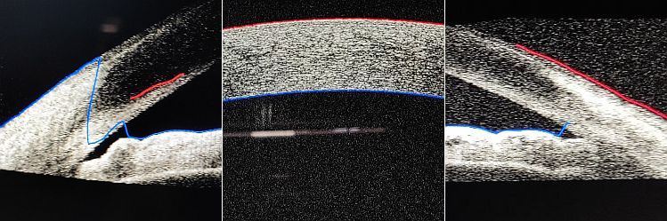
Anterior drainage angle imaging;
This is relevant to more serious acute angle closure glaucoma cases.
-
- occludable drainage angle anatomy (likely to give rise to angle closure glaucoma attack) may require iridotomy. (specialist surgical laser intervention)
-
- non-occludable drainage angle anatomy (unlikely to give rise to angle closure glaucoma attack) which is not requiring surgical intervention.
-